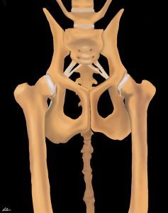
Hip Dysplasia (HD) is a type of developmental orthopedic disease and a common cause of lameness in dogs. Hip Dysplasia is a condition that dogs are born with that eventually leads to arthritis in the hip.
Hip Dysplasia Overview
The word “dysplasia” means abnormal growth and development. The hip joint is where the thigh bone, or femur, meets the pelvis. This joint is shaped like a ball (femoral head) and socket (acetabulum of the pelvis).
Hip dysplasia is the abnormal development of the hip joint. This results in looseness of the joint due to decreased coverage of the femoral head by the acetabulum. Whenever a young dog with HD walks, the femoral head will slip in and out of the joint; this causes pain and wearing away of cartilage in the joint. Over time, the abnormal motion in the joint results in arthritis.
What Dogs Will Develop Hip Dysplasia?
Hip dysplasia is genetic, meaning that dogs are born predisposed to develop the condition. But, there are a number of factors that can influence the severity and time when symptoms develop. These factors include:
- Weight
- Body condition
- If the dog is spayed or neutered
- Their age at the time of spay or neuter
- Age
- Body conformation
A study of genetically predisposed Labradors found that the development and progression of HD were significantly delayed or decreased in dogs that maintained a lean body condition through caloric restriction. These dogs were “on the skinny side of normal” for their entire life.
When compared to dogs that were 15-20% heavier, the lean dogs developed signs of arthritis at 12 years of age whereas the overweight dogs showed signs at 6 years. The lean dogs also lived 2-3 years longer!
Diagnosis
Dogs with HD often show clinical signs at two points in time:
- 4 months to 3-4 years old
- >7 years of age
Puppies and young dogs with HD will be less active, be reluctant to play, jump or climb stairs, might have a bunny hopping gait, and may seem under-developed in the back legs in terms of muscle mass. They might also vocalize pain, though this is very rare.
As early as 16 weeks of age, your veterinarian can make a diagnosis by palpating the hips to check for hip looseness (a test called Ortolani). A special type of x-rays called PennHIP can show the degree of hip looseness and the likelihood of developing arthritis symptoms. The traditional x-ray view may or may not show hip dysplasia.
HD leads to the development of arthritis over months and years. Mature dogs with hip arthritis show varying degrees of lameness that is worse after rest and heavy exercise, reluctance to jump or go upstairs, hind limb muscle atrophy, and decreased interest in walks and other activities.
The severity of HD and associated arthritis can be assessed with x-rays, but there is not a direct correlation between the severity shown on the x-ray and clinical symptoms. In other words, x-rays may show severe degenerative changes to the joint, but your dog might hardly show any symptoms.
Treating Hip Dysplasia
Non-surgical management: A diagnosis of HD and arthritis does not mean your dog will need surgery. In fact, most dogs with HD and arthritis respond well to a multi-tiered approach that includes
- Maintaining a lean body condition
- Modifying the type of activity they do while ensuring regular, low impact exercise
- Making changes to their environment
- Managing pain
- Physical rehabilitation
- Joint injections
- Joint supplements
- Complementary medicine, such as acupuncture
Surgical Treatment
There are 4 types of surgical treatment for hip dysplasia.
Two are performed in juvenile dogs with the goal of preventing arthritis, and two are typically performed in adult dogs that already have arthritis symptoms.
Surgeries to Prevent Arthritis (Juvenile)
Juvenile Pubic Symphysiodesis (JPS):
This is a relatively simple procedure that involves closing one of the growth plates in the pelvis to alter the growth of the acetabulum so that the femoral head is more fully covered.
This procedure can be effective for dogs with mild to moderate HD as long as it is performed in puppies between 16-20 weeks of age. Studies have shown that the development of the pelvis is improved and less arthritis can be expected long term.
Triple or Double Pelvic Osteotomy (TPO/DPO):
This surgery involves cutting the pelvis in 2 or 3 places and rotating the acetabulum to provide better coverage of the femoral head.
This is a much more invasive procedure than the JPS but the goal is the same. It is performed in puppies typically between 6-8 months of age. It can be effective in reducing the progression of arthritis but only if it is performed prior to the onset of arthritis.
Surgeries to Treat Arthritis Related to HD (Adult)
Treating arthritis secondary to HD involves either replacing the joint or removing the articulation and bone-on-bone contact. These surgeries are reserved for when non-surgical management efforts have been exhausted.
Total Hip Replacement (THR):
This is the gold standard surgery for HD, and results can be excellent with dogs returning to very active lives. However, there are potential risks associated with surgery. If non-surgical treatment options have not been successful with your dog, consider consulting a surgeon to discuss post-operative care and potential risks.
Femoral Head and Neck Ostectomy (FHO):
This procedure removes the femoral head and neck, which stops the bone-on-bone contact of the hip and decreases pain. Many dogs do very well with this procedure and can return to a very high level of activity with reduced pain. Post-operative rehabilitation soon after surgery is very important, especially for large dogs.
Illustrations by Stephanie Erdesz


