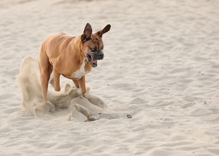
Elbow Dysplasia (ED) is a type of developmental orthopedic disease and a common cause of lameness in growing dogs. It affects the joints and eventually leads to arthritis in the elbow.
Elbow Dysplasia Overview
Elbow Dysplasia is defined as abnormal development of the elbow joint.
Three individual conditions are recognized as forms of ED:
- Fragmented Medial Coronoid Process (FMCP)
- Osteochondrosis/Osteochondritis dessicans (OC/OCD)
- Ununited Anconeal Process (UAP)
Affected dogs may have dysplasia in one elbow or both, and may have more than one type of pathology in the same elbow joint. All of these conditions will lead to arthritis in the joint.
Elbow dysplasia is considered a genetic condition but dietary factors and trauma may play a role in certain cases. Similar to hip dysplasia, factors such as body weight/ condition and activity level can influence the development and progression of symptoms.
FRAGMENTED MEDIAL CORONOID PROCESS (FMCP)
What is FMCP?
FMCP is the most common pathology present in cases of elbow dysplasia. It is characterized by fissures or complete separation of the medial coronoid process from the ulna. This may result from abnormal stresses placed on the coronoid process during development.
What are the clinical signs and how is FMCP diagnosed?
Common clinical signs of FMCP begin between 5-8 months of age and include acute or chronic intermittent lameness, stiffness, and stilted gait. Lameness is usually intensified by exercise and is prominent after resting. In some cases, lameness is not apparent until later in life. Palpation by the vet may show joint swelling, and pain upon extension and flexion of the leg. Older dogs may have crepitus (crackling) within the joint.
The clinical signs of FMCP are very similar to those of OCD, and it is likely that the two coexist in a third of cases. The majority of cases are bilateral, but clinical signs may be unilateral.
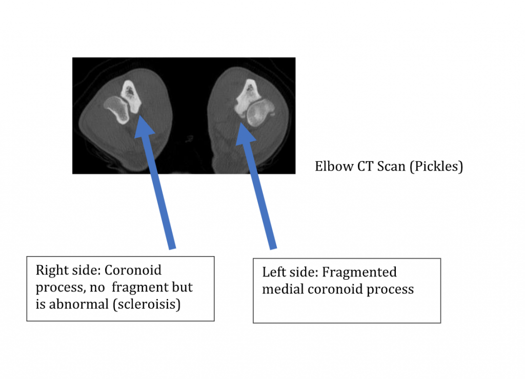
Radiographs may show degenerative changes in the elbow, including sclerosis (increased whiteness of the bone) and osteophyte (bony spur) formation. It is difficult to visualize the coronoid process on radiographs, and FMCPs are rarely diagnosed on radiographs alone.
Definitive diagnosis requires either arthroscopic joint exploration or CT scans.
What is the recommended treatment for FMCP?
Treatment consists of removal of the FMCP and any other cartilage fragments through arthroscopic or traditional open surgery.
However, it is important to realize that while removing the fragment often provides improvement in lameness, this will not be a curative surgery and long term management of arthritis will be required.
OSTEOCHONDRITIS DESSICANS (OCD)
What is OCD?
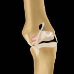
Osteochondrosis is a defect in bone growth that results in soft lesions in the cartilage. As lesions progressively worsen with activity, cracks and fissures form, and eventually, the cartilage may partially split away, forming a cartilage flap which causes pain when it moves in the joint space. This condition is termed osteochondritis dessicans (OCD).
Once formed, this cartilage flap cannot heal back down to the underlying lesion bed. Instead, it will either continue to degenerate in place or may break free and migrate to another area of the joint (these free pieces are known as “joint mice”). In either case, the presence of the flap will cause pain and inflammation within the joint. OCD not only occurs in the elbow but can be seen in any joint.
What are the clinical signs and how is OCD diagnosed?
Common clinical signs of OCD begin between 5-8 months of age and include acute or chronic intermittent lameness, stiffness, and stilted gait. Lameness is usually intensified by exercise and is prominent after resting.
In some cases, lameness is not apparent until later in life. Palpation by the vet may show joint swelling, and pain upon extension and flexion of the leg. Older dogs may have crepitus (crackling) within the joint.
Diagnosis is confirmed by visualizing the OCD lesion, flap or joint mice on Radiographs, CT and/or during joint exploration.
What is the recommended treatment for OCD?
Treatment for OCD consists of surgical removal of the flap and any other loose cartilage or joint mice, and smoothing out the area of the lesion to stimulate the filling-in of the lesion area with fibrocartilage (scar-tissue-type cartilage). This may be accomplished arthroscopically or through traditional open surgery.
However, it is important to realize that while surgery often provides improvement in lameness, this will not be a curative surgery and long term management of arthritis will be required.
UNUNITED ANCONEAL PROCESS (UAP)
What is UAP?
UAP is characterized by the failure of the growth center of the anconeal process to fuse properly with the olecranon. This leads to instability or detachment of the process and osteoarthritis of the elbow.
It is most commonly diagnosed in German Shepherds.
What are the clinical signs and how is UAP diagnosed?
Clinical signs are usually not seen before 5-8 months of age. Occasionally, lameness is not observed until the dog is several years old. In the earliest stages, the only clinical signs may be a slight limp, swelling on the outside of the elbow and standing or walking with the paw turned out. Palpation of the joint in older dogs may reveal swelling and crepitus (crackling) within the joint.
A preliminary diagnosis may be made by clinical signs, age, and breed. The diagnosis will be confirmed by Radiographs showing the partially or fully detached anconeal process, as well as possible osteophytes (bony spurs) throughout the joint.
What is the recommended treatment for UAP?
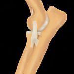
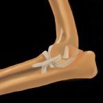
There are several surgical options that may be considered for young dogs diagnosed with UAP. These include:
- arthroscopy to evaluate the entire joint
- reattaching the anconeal process with a screw
- cutting the ulna bone to relieve tension on the UAP so that it may fuse,
- surgery to remove the anconeal process.
Surgical options will be discussed by your veterinary surgeon. Like other components of elbow dysplasia, UAP will likely result in some degree of long-term arthritis.
Elbow Dysplasia: Treatment
Non-surgical
All forms of elbow dysplasia will result in arthritis, even with early surgical intervention.
If your dog has elbow dysplasia, a long-term, multi-pronged approach to managing arthritis will be needed. That includes:
- Pain management
- Activity modification
- Environmental adaptations
- Weight management
- Rehabilitation techniques
- Intra-articular therapies
- Read the story of Pickles, a crisis response dog with elbow dysplasia
Surgical Treatment of Elbow Arthritis
Unlike hip dysplasia, joint replacement for elbow arthritis is not a highly successful procedure.
There are surgeons continuing to work on refining the implants and surgical technique, but to date, total elbow replacement is not widely performed and carries a large number of risks.
Other surgical options for elbow arthritis in mature dogs include canine unicompartmental elbow replacement (CUE) and Sliding Humeral Osteotomy (SHO); these procedures are also not widely performed and have shown mixed results in clinical patients.
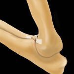
Illustrations by Stephanie Erdesz