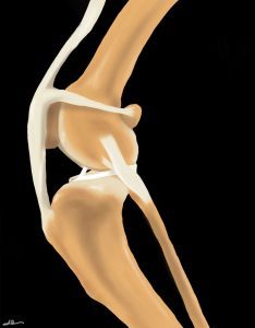
Cranial cruciate ligament (CCL) disease, also referred to as CCL rupture, is among the most common causes for pelvic limb lameness in dogs and should be ruled out prior to treating a dog for simply presumed hip arthritis.
See this article for additional information on what the CCL is and why it tears.
Treatment of CCL disease (sprain/tear/rupture) can be surgical or non-surgical. Unfortunately, the CCL will not heal on its own. Over time, the injury typically progresses to a complete tear. The body’s natural reaction to the joint being unstable is to lay down scar tissue to stabilize the knee. This takes many months to years. Meanwhile, arthritis is progressing rapidly. With or without surgery, arthritis management techniques will be needed to optimize your dog’s comfort and activity.
While surgery is not an option for all dogs, it is the most effective and reliable means of returning dogs to a high level of function and minimizing the progression of arthritis.
Surgery
The first part of surgery is exploration of the stifle joint in order to evaluate the CCL and the medial and lateral menisci. Joint exploration may be performed by arthrotomy (small opening in the joint to visualize the structures) or arthroscopy (inserting a camera into the joint). In cases of meniscal tear, the torn portion of the meniscus will be removed. If the medial meniscus cannot be fully evaluated, a meniscal release procedure may be performed. This involves cutting the attachment of the meniscus on the tibia to allow it to fall away from the compressive force of the femur, thereby reducing the chance of future tears and future surgery.
After evaluating the CCL and meniscus, a technique will be performed to help stabilize the stifle joint. Surgical stabilization is divided into two categories: osteotomy and extra-capsular techniques.
Osteotomies (TPLO, TTA, CBLO, CWO)

Osteotomy means “to cut the bone.” Osteotomy techniques include tibial plateau leveling osteotomy (TPLO), tibial tuberosity advancement (TTA), CORA based leveling osteotomy (CBLO), and closing wedge osteotomy (CWO). These techniques all aim to change the biomechanical forces acting on the stifle joint in order to eliminate the “rolling off the hill” motion, (called cranial tibial translation or cranial tibial thrust), that occurs with partial or complete CCL rupture.
In cases of Grade 1 sprain or very partial CCL rupture, these surgeries may decrease the stress on the CCL and decrease the pain that occurs when the injured ligament is stretched.
Of the osteotomy procedures, TPLO is the most well studied and has the most research to support its efficacy. While all surgeries carry a risk of complication, the TPLO has a very high success rate when performed by experienced surgeons. Other osteotomy techniques may also provide excellent outcomes, but results will be closely linked to the experience of the surgeon. Ultimately, the best surgery will be the one the surgeon feels most comfortable with and recommends for your dog.
This is a great website to learn more about the TPLO: www.TPLOinfo.com
TPLO
Technique
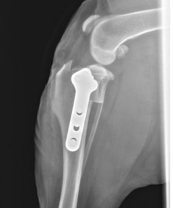
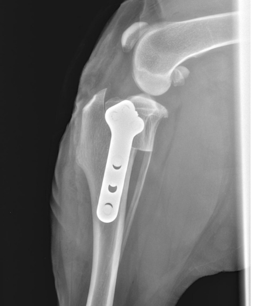
The theory for the technique is based on the fact that the slope of the tibial plateau causes cranial tibial thrust (instability) in the CCL deficient stifle (either the CCL isn’t working fully or is completely ruptured). The surgery produces functional stability of the knee by leveling or flattening of the tibial plateau slope. This is accomplished by cutting the top portion of the tibia (osteotomy) and leveling the tibial plateau to prevent cranial tibial thrust during weight-bearing. The tibia is then stabilized with a bone plate and screws.
Prognosis
The TPLO has gained worldwide acceptance by providing dogs with a faster recovery, better functional outcome, and decreased development of osteoarthritis as compared to dogs treated with other procedures. Over the last 20+ years, the TPLO has been the most commonly performed surgery for CCL rupture in dogs. The typical dog will achieve about 95% of normal limb function following TPLO surgery. However, the level of pre-existing arthritis can also affect the prognosis. Dogs with severe arthritis may have persistent lameness.
Complications
The following complications have been reported:
- Infection
- Inflammation of the patellar tendon
- Fracture of the tibial tubercle and patella
- Breakage or loosening of the bone plate or screws
- Delayed healing of the osteotomy site
- Rupture of the caudal cruciate ligament
- Post-operative meniscal injury
- Bone cancer (very rare).
Many of these complications can be the result of too much post-operative activity. When performed by experienced, board-certified surgeons, the risk of complications with TPLO is typically less than 5%.
Recovery
One of the true benefits of this procedure is that it relies on bone healing, which is very predictable. The osteotomy should heal in 8-12 weeks, and once healed, the bone will be as strong as it was before surgery.
However, during the healing process, it is critical that excessive force is not placed on the bone. No running, jumping, off-leash activity or play is allowed until the bone has healed. Dogs must begin using their leg within the first 1-2 weeks after surgery in order to stimulate healing and build muscle, and then they should have progressively longer leash walks over the recovery time.
Additional rehabilitation therapy can help speed muscle strengthening and guide return to activity. Most dogs with a TPLO are expected to be using the leg with near-normal function by 10-12 weeks post-op.
Extra-capsular Suture/ Lateral Suture/ Tight-Rope
This technique uses strong sutures (fishing line or TightRope) placed just on the outside of the joint capsule to mimic the path of the CCL. Eventually, scar tissue aligns itself around the sutures and ultimately it is this scar tissue that provides stability to the stifle. This technique is typically performed in small breed dogs (less than 15 pounds) and occasionally added to a TPLO for additional stability.
Prognosis
Extracapsular repairs rely on scar tissue to ultimately stabilize the stifle. Scar tissue is somewhat unpredictable in the time it will take to form and the ultimate strength it can provide. The prognosis following extracapsular surgery is generally fair to good for smaller dogs; however, larger dogs will often stretch out the extracapsular sutures and have continued instability within the joint. This will result in decreased range-of-motion (stiffness) and progression of arthritis (although likely much slower than without surgery).
Smaller dogs would be expected to return to about 90-95% of normal limb function, while larger dogs may only obtain 75% normal limb function. Post-operative rehabilitation has been shown to improve outcomes following extracapsular repair. The level of pre-existing arthritis can also affect the prognosis. Dogs with severe arthritis may have persistent lameness.
Complications
As with any surgery, complications are a possibility. Although not common, the following have been documented:
- Infection, reaction or irritation to the sutures
- Breakage of the stabilization sutures before the development of sufficient scar tissue,
- Post-operative damage to the meniscus.
Some type of complication occurs in about 5-10% of dogs, but the complication rate can be much higher in larger-breed dogs.
CCL Non-Surgical Management
While surgery is generally recommended as the first-line option for treating CCL disease, there will be cases in which surgery is not the right option for the dog. The expected course without any surgical intervention is that the dog will develop progressive thickening/scarring/fibrosis around the stifle; this is the body’s attempt to provide stability. This process takes months to years, and progression of arthritis, including muscle atrophy, will occur.
Non-surgical management should include comprehensive pain management and possibly physical rehabilitation (Find a rehab vet here). The goals of rehabilitation include:
- Reducing inflammation and pain
- Supporting joint health
- Increasing muscle strength
- Weight loss (if indicated)
- Supporting the body while fibrosis develops around the stifle
Non-surgical management of CCL disease with rehabilitation is less predictable in the time course of returning to activity, and full/ normal function is not expected. Rather, some degree of lameness should be expected. The degree and frequency of lameness will depend on the degree of CCL rupture (partial vs. complete), concurrent meniscal pathology and degree of arthritis. One study suggests that 60% of dogs with conservative management for CCL disease can have an acceptable outcome. Dogs in this study were placed on a weight loss plan, received NSAIDs, and participated in at least 6 sessions of formal rehabilitation.
One thing to note with non-surgical management is that the CCL is not expected to heal. Improvements seen over months and years are generally due to the natural development of per-articular fibrosis (scar tissue around the knee).
Read more about non-surgical management here.
Article written by Dr. Kristin Shaw
Reviewed/updated 12/2024
References
Slocum B, Slocum T. Tibial plateau leveling osteotomy for repair of cranial cruciate ligament rupture in the canine. Vet Clin N Am-Small 23:777–95, 1993
Hoffmann DE, Miller JM, Ober CP, et al: Tibial tuberosity advancement in 65 canine stifles. Vet Comp Orthop Traumatol 19:219–227, 2006
Kim SE, Pozzi A, Kowaleski MP, et al: Tibial osteotomies for cranial cruciate ligament insufficiency in dogs. Vet Surg 37:111–125, 2008
Raske M, Hulse D, Beale B, et al: Stabilization of the CORA based leveling osteotomy for treatment of cranial cruciate ligament injury using a bone plate augmented with a headless compression screw. Vet Surg 42: 759-764, 2013.
Au, KK, Gordon-Evans WJ, Dunning D: Comparison of short‐and long‐term function and radiographic osteoarthrosis in dogs after postoperative physical rehabilitation and tibial plateau leveling osteotomy or lateral fabellar suture stabilization. Vet Surg 39:173-180, 2010
Nelson SA, Krotscheck U, Rawlinson J, et al. Long-term functional outcome of tibial plateau leveling osteotomy versus extracapsular repair in a heterogenous population of dogs. Vet Surg 42:38-50, 2013
Christopher SA, Beetem J, Cook JL. Comparison of long-term outcomes associated with three surgical techniques for treatment of cranial cruciate ligament disease in dogs. Vet Surg 42:329-334, 2013
Lazar TP, Berry CR, Dehaan JJ, et al: Long‐term radiographic comparison of tibial plateau leveling osteotomy versus extracapsular stabilization for cranial cruciate ligament rupture in the dog. Vet Surg 34:133–141, 2005
Gordon-Evans WJ, Griffon DJ, Bubb C, et al: Comparison of lateral fabellar suture and tibial plateau leveling osteotomy techniques for treatment of dogs with cranial cruciate ligament disease. J Am Vet Med Assoc 243:675-680, 2013
Thieman K M, Tomlinson J L, Fox D B, Cook C, Cook J L. Effect of meniscal release on rate of subsequent meniscal tears and owner assessed outcome in dogs with cruciate disease treated with tibial plateau leveling osteotomy. Vet Surg. 2006 Dec;35(8):705-10.
Cook J, Luther J, Beetem J, et al: Clinical comparison of a novel extracapsular stabilization procedure and tibial plateau leveling osteotomy for treatment of cranial cruciate ligament deficiency in dogs. Vet Surg 2010;39(3):315-23
Boudrieau RJ. Tibial plateau leveling osteotomy or tibial tuberosity advancement? Vet Surg. 2009 Jan;38(1):1-22.
Lazar TP, Berry CR, deHaan JJ, Peck JN, Correa M. Long-term radiographic comparison of tibial plateau leveling osteotomy versus extracapsular stabilization for cranial cruciate ligament rupture in the dog. Vet Surg. 2005 Mar-Apr;34(2):133-41
Priddy NH 2nd, Tomlinson JL, Dodam JR, Hornbostel JE. Complications with and owner assessment of the outcome of tibial plateau leveling osteotomy for treatment of cranial cruciate ligament rupture in dogs: 193 cases (1997-2001). J Am Vet Med Assoc. 2003 Jun 15;222(12):1726-32.
Lafaver S, Miller NA, Stubbs WP, Taylor RA, Boudrieau RJ. Tibial tuberosity advancement for stabilization of the canine cranial cruciate ligament-deficient stifle joint: surgical technique, early results, and complications in 101 dogs. Vet Surg. 2007 Aug;36(6):573-86.
Stein S, Schmoekel H. Short-term and eight to 12 months results of a tibial tuberosity advancement as treatment of canine cranial cruciate ligament damage. J
Small Anim Pract. 2008 Aug;49(8):398-404.
Marsolais, GS, Dvorak G, Conzemius MG: Effects of postoperative rehabilitation on limb function after cranial cruciate ligament repair in dogs. J Am Vet Med Assoc 220:1325-1330, 2002
Monk, ML, Preston CA, McGowan CM: Effects of early intensive postoperative physiotherapy on limb function after tibial plateau leveling osteotomy in dogs with deficiency of the cranial cruciate ligament. Am J Vet Res 67:529-536, 2006
Romano LS, Cook JL: Safety and functional outcomes associated with short-term rehabilitation therapy in the post-operative management of tibial plateau leveling osteotomy. Can Vet J 56:942-946, 2015
Baltzer WI, Smith-Ostrin S, Ruaux CG: Evaluation of the clinical effects of diet and physical rehabilitation in dogs following tibial plateau leveling osteotomy. J Am Vet Med Assoc 252:686-700, 2018
Verpaalen VD, Baltzer WI, Smith-Ostrin S, et al: Assessment of the effects of diet and physical rehabilitation on radiographic findings and markers of synovial inflammation in dogs following tibial plateau leveling osteotomy. J Am Vet Med Assoc 252:701-709, 2018
Wucherer KL, Conzemius MG, Evans R et al: Short-term and long-term outcomes for overweight dogs with cranial cruciate ligament rupture treated surgically or nonsurgically. J Am Vet Med Assoc 2013;242:1364-1372
Hart JL, May KD, Kieves NR, et al: Comparison of owner satisfaction between stifle joint orthoses and tibial plateau leveling osteotomy for the management of cranial cruciate ligament disease in dogs. J Am Vet Med Assoc 2016;249(4):391-398

