guest post by Laurie McCauley, DVM, DACVSMR, CCRT, CVA, CVC
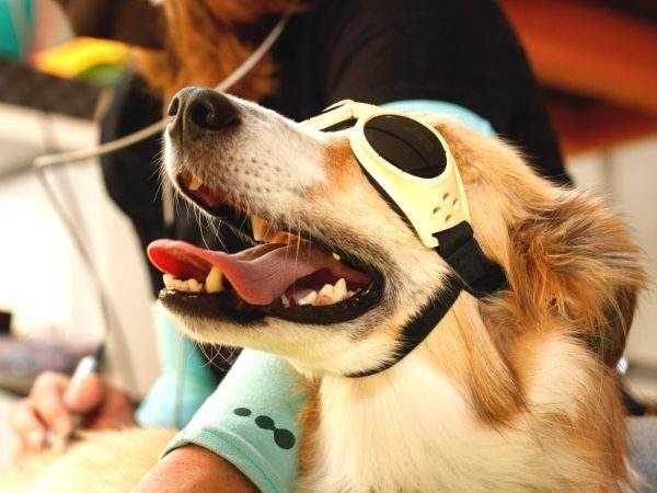
When should you use laser therapy with your OA patients?
In a way, “When should I use laser therapy for a patient with osteoarthritis?” is a trick question. This is because the answer is ideally, before, during, and after the onset of osteoarthritis (OA), which most often causes the clinical sign of lameness. Let’s start with some definitions of osteoarthritis, as well as its progression to osteoarthrosis.
Osteoarthritis is:
- an inflammatory condition of the joint which, in canine and feline patients, is often due to secondary causes (trauma, joint incongruity, over-use), which, if untreated, often results in varying degrees of progressive cartilage degeneration and secondary soft tissue and bone changes.
Osteoarthrosis is:
- the sequela to chronic, longstanding osteoarthritis when it has progressed past the point of inflammation to non-inflammatory condition.
- the result of progressive degenerative changes in the cartilage of one or more joints accompanied by thickening and overgrowth of adjacent bone and that is identified symptomatically by stiffness, swelling, pain, deformation of joints, and loss of range of motion.
In other words, we can treat the inflammation of the joint with the goal of slowing down the progression that will lead to osteoarthrosis, the degenerative changes that we frequently see, and that are not responsive to NSAIDs. We can still treat these joints with the goal of reducing inflammation even if they have chronic osteoarthrosis if there is acute inflammation on top of their chronic osteoarthrosis. An example would be the dog that has known osteoarthrosis and then comes up lame after playing in the dog park or after a run with their owner.
 What joints are good candidates for laser?
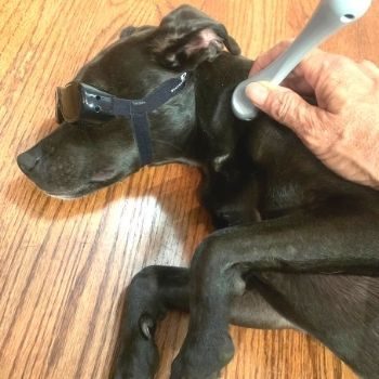
Going back to early in the process, I consider any joint with any crepitus or effusion a candidate for laser therapy, even if we don’t have pain and stiffness as a clinical sign yet. Why wait until the cartilage damage is done if we can slow the progression down or stop it from having irreversible changes? Inflammation and stiffness can be alleviated once we have bony change, but at this point, we are fighting the sequela rather than preventing the process.
Osteophytes form where the periosteum meets the cartilage, at the line of the joint capsule. Knowing where that line is located allows us to treat that tissue directly, with an appropriate amount of energy. That amount of energy depends on the depth of the tissue and the wavelength of the laser.
For instance, treating the hip joint capsule will require treating with a specific angle and significantly more energy than if we were treating a stifle joint capsule that is comparatively superficial. We would also treat with significantly more energy if the wavelength of our laser is 980 nm versus 904 or 830 nm.
With a laser with a 980nm wavelength, laser energy is absorbed by water significantly more than the other common wavelengths and so more energy is used to saturate the tissue to allow increased penetration. The absorption by water is why there is heat created, energy stays in the superficial tissue until it is saturated and then it passes through. Lasers that are in the 800nm wavelength range have significantly less absorption by water but have more absorption by melanin and the energy needs to be delivered slower to prevent heating at the skin level. Lasers with a 904 or 905nm wavelength have the most penetration as they have less absorption by water and melanin. They also are usually super-pulsed, which means they are delivered with high amounts of energy, 25-100W, but over a very short amount of time with rest periods between so as not to create heat.
When treating osteoarthritis or trying to prevent the clinical signs, the goals are to treat the joint capsule to decrease active inflammation if it is present, reduce local pain, treat the cartilage surface that is accessible to have a positive effect on the chondrocytes and enhance strengthening of the muscles, tendons, and ligaments that help stabilize the joint. The cells that produce the inflammatory mediators that affect the joint are positioned on the internal surface of the joint capsule.
Benefits of laser
Laser has been shown to reduce interleukin 1, 6 (IL- 1, IL-6), Tumor Necrosis Factor (TNF), and cyclooxygenase-2 (COX-2) release, thereby reducing the active inflammation that leads to added degenerative changes to the cartilage. Laser has also been shown to increase endogenous opioid production and enhance the production of reactive oxygen species which then activate transient receptor potential vanilloid 1 (TRPV1) ion channels, both of which decrease pain.
High doses of laser energy can lower nociceptor conduction velocity, temporarily break up the microtubules in the axons that transport the mitochondria down the length of the axon, and reduce mitochondrial respiration. These actions decrease ATP and as well as action potentials received at the dorsal horn interneuron, leading to a reduced perception of pain to the brain. At low doses, laser has been shown to increase fibroblast for healing, enhance angiogenesis, accelerate healing of tendon and ligament injuries, improve cartilage thickness in patients with OA, and protect articular cartilage. There are nociceptive pain receptors in the joint capsule as well as in the tendons and ligaments that surround the joint that should be targeted to have maximum therapeutic effect. The positioning of the joint is important to be able to have an effect on the internal components of the joint, the chondrocytes, menisci, and ligaments, as well as the more superficial components of the joint capsule, muscle, and tendon.
Positioning the joints
When we think about joint positioning for optimum effect on the chondrocytes, we first consider that the joint should be non-weight bearing or offloaded, as penetration is significantly reduced if the laser energy has to penetrate the bone, versus penetrating the joint capsule and synovial fluid, to reach the chondrocytes. Traction, varus, valgus, flexion, or extension should be performed depending on which joint surface you are treating.
Knowing the anatomy to know what cartilage surfaces you can access and how to best position the probe and the joint for optimal effect is imperative. The anatomy of each joint should be reviewed and treated appropriately. Most joints have a medial and lateral collateral ligament that play a major role in preventing medial and lateral sheer force, as well as some role in rotation. These are usually easy to find, making position less important, but with other ligaments like the cruciate ligament, position plays a significant role in the ability to access it.
Knowing the location of the menisci and the best angle to have optimum penetration are considerations worth having in the case of cruciate disease, or even inflammation of the stifle, for maximum support of the local tissue. To access the cruciate ligaments, the best position is when the stifle is in moderate extension. The goals for treating these structures include improving blood supply and potentially increasing the ATP in the cells that lay down the collagen that keeps the ligaments strong. The best positioning to treat the joint capsule for each joint could be a paper in itself and is detailed in the Optimum Pet Vitality “Optimum Laser Therapy” course found at Optimum Laser Therapy.
What to treat with laser?
The next piece of the puzzle is knowing which muscles, tendons, and ligaments to treat with laser as the goal is to treat the structures that stabilize the affected joint. This is particularly important if the cause of the inflammation and secondary osteoarthritis is laxity of the joint as in hip dysplasia. This is just as important along the spine as it is in the appendicular joints. It is also important to note if pain has led to the lack of muscle engagement, leading to atrophy of the supporting structures. If this is the case, resolving pain is our first goal; and then improving the strength of the supporting structures and stimulating healing of the affected tissue is our second goal. Different laser settings/variables are chosen for each goal.
Once we know which structures are involved, we can laser the muscles before exercises to increase the rate of strengthening and improve blood flow to the tendons, indirectly via the muscles. In the case when the tendons have a paratendinous sheath that supplies the nutrients instead of nutrients being supplied from blood flow from the muscle, lasering the sheath that surrounds the tendons is essential. Lasering the supporting ligaments also directly improves blood flow to these structures. As an example, we know the quadriceps and gastrocnemius muscles aid in preventing a sheer force at the stifle that would stress the cranial cruciate ligament. Therefore, anytime we are dealing with cruciate disease or trying to prevent a cruciate from rupturing, as in a dog with straight rear limb confirmation, it makes sense to laser these muscles before exercises are performed.
Knowing where and how to laser patients with OA is analogous to having a knowledge of which antibiotic works best in certain tissues as well as performing a culture and sensitivity to treat said infection. Not knowing where and how to laser patients with OA is analogous to thinking there is an infection in your patient and treating the presumed infection by reaching up on a shelf and picking an antibiotic randomly. It may work great, it may work some, and it may not work at all. And it is not the antibiotic’s fault.
Improving the prescriber’s knowledge improves outcomes.
Want to learn more?
For more information on understanding laser therapy and how to gain Optimum results, check out Optimum Laser Therapy, a thorough course that will help you have optimum results with all of your laser patients.
References
Alves, A.C., Vieira, R., Leal-Junior, E., Dos Santo, S, S., Ligeiro, A.P., Albertini, R., Junior, J. & De Carvalho, P. 2013. Effect of low-level laser therapy on the expression of inflammatory mediators and on neutrophils and macrophages in acute joint inflammation. Arthritis Res Ther, 15(5), R116.
Assis, L., Manis, C., Fernandes, K. R., Cabral, D., Magri, A., Veronez, S. & Renno, A. C. 2016. Investigation of the comparative effects of red and infrared laser therapy on skeletal muscle repair in diabetic rats. Am J Phys Med Rehabil, 95, 525-34.
Bublitz, C., Medalha, C., Oliveira, P., Assis, L., Milares, L. P., Fernandes, K. R., Tim, C. R., Vasilceac, F. A., Mattiello, S. M. & Renno, A. C. 2014. Low-level laser therapy prevents degenerative morphological changes in an experimental model of anterior cruciate ligament transection in rats. Lasers Med Sci, 29, 1669-78.
https://www.arthritis.org/diseases/osteoarthritis
Passarella et al 1984. Increase of protein electrochemical potential and ATP synthesis in rat liver mitochondria irradiated in vitro by helium neon laser. FEBS Lett, 175(1), 95-99.
Karu 1988. Molecular mechanisms of the therapeutic effects of low intensity laser radiation. Lasers in the life sciences,2, 53-84.
Nasu et al 1989. Cytochemical effects of GaAlAs diode laser radiation on rat saphenous artery calcium ion dependent adenosine triphosphatase activity. Laser therapy, 1(2), 89-92.
Nicholas T. Kruse, Christopher R. Silette, and Barry W. Scheuermann. Influence of passive stretch on muscle blood flow, oxygenation and central cardiovascular responses in healthy young males. Integrative Cardiovascular Physiology and Pathophysiology. 01 MAY 2016https://doi.org/10.1152/ajpheart.00732.2015
S, G. N., Kamal, W., George, J. & Manssor, E. 2016. Radiological and biochemical effects (CTX-II, MMP-3, 8 and 13) of low-level laser therapy (LLLT) in chronic osteoarthritis in Al-Kharj, Saudi Arabia. Lasers Med Sci. 32(2), 297-303.
Sakurai Y, Yamaguchi M And Abiko Y. 2000. Inhibitory effect of low-level laser irradiation on LPS-stimulated prostaglandin E production and cyclooxygenase-2 in human gingival fibroblasts. European Journal of Oral Science 108: 29-34.