by Kara Amstutz, DVM, CCRT, CVPP, CVA
Veterinarians and pet owners alike face a sometimes elusive condition that causes pain in our dog companions which can take a toll on daily mobility, ability to work or play, and overall quality of life.
The problem arises at the lumbosacral (LS) joint, a transition zone in the spine where the last lumbar vertebrae meet the sacrum. This particular joint is at risk for degenerative changes because the force being generated by the hind limbs to propel the dog forward has to travel up through this area. This causes excess flexion and extension of the LS joint and, over time, can lead to disc disease such as bulging discs, compression on nerve roots (often leading to sciatica pain), new bone formation called spondylosis, and osteoarthritis in the spine. The body’s natural response is to stabilize this joint and remodel the tissues, however, this gives rise to a dynamic compression of the nerve roots in the canal and as they exit the spine. This means extreme pain can occur in certain positions of the spine – most likely in extension of the lumbosacral joint.
We typically see DLSS in middle age to older dogs as it takes time for the degenerative changes to occur and cause clinical symptoms. The condition can occur in any breed, however, the most likely breed to develop DLSS is the German Shepherd, which can show symptoms as early as 15 months of age. The higher incidence and young onset of clinical signs are secondary to the fact GSDs have a higher chance of being born with conformational abnormalities that lead to early-onset DLSS. Breeding pairs in predisposed breeds should be evaluated for these conformational abnormalities and removed from the breeding program if identified.
We also know working dogs have a higher prevalence of clinical signs. Partly, this is due to the breeds chosen to be working dogs, but it is also related to the extreme conditions they put their bodies through in a normal working day. A dog that has the luxury of lounging on the couch all day and going for a couple of easy walks each day can hide symptoms of DLSS much better than a working dog that is expected to search high, chase a suspect, or jump out of a vehicle many times per day.
The most common symptoms a pet owner will notice include
- low back pain
- hesitation to jump up
- slow to rise from a down position
- difficulty with stairs
- difficulty maintaining posture for elimination
- low tail carriage
- and intermittent lameness.
A recent study found that behavioral changes such as refusal to do certain commands (jump up, sit), appetite loss, and self-mutilation of the tail can occur in working dogs. This can be catastrophic for a working dog’s career. In fact, DLSS is, unfortunately, the third most common reason for humane euthanasia in working dogs.
Source: Meij, B, Degenerative lumbosacral stenosis in Dogs. The Veterinary clinics of North America, Small Animal Practice, 2010
In addition to pain, if there is enough compression of the spinal nerve roots within the vertebral canal, or as they exit the spinal column, neurologic deficits may be observed. Typical neurologic changes include weakness on one or both hind limbs, dragging the paw or knuckling, worn toe nails, hesitation to put the limb down to bear weight (described as nerve root signature), bladder and bowel control issues, and abnormal reflexes.
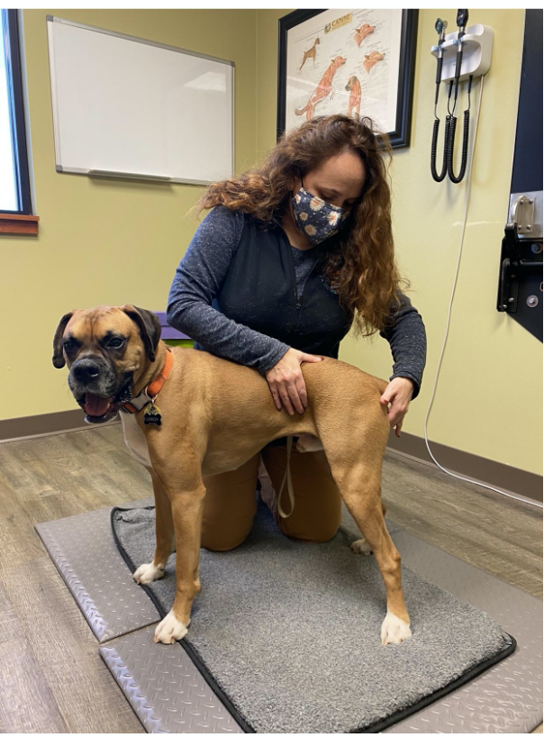
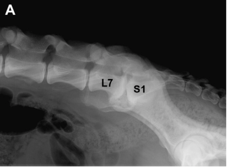
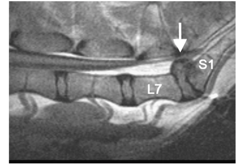
A complete orthopedic and neurologic exam performed by an experienced veterinarian or canine rehabilitation therapist can lead to the suspicion of DLSS. There are special tests that can help localize pain and dysfunction to the LS area. However, imaging of the region is needed to confirm the diagnosis. Radiographs may reveal spondylosis at the LS joint, narrowing of the disc space, as well as osteoarthritis. Radiographs can also demonstrate additional pathology that is often associated with DLSS such as hip dysplasia and spine conformational abnormalities. The gold standard for evaluating the spine and nerve roots is utilizing Magnetic Resonance Imaging (MRI). This allows for localizing any spine or root compression which is vital if surgical intervention is necessary.
Treating DLSS patients typically begins with conservative therapies for most pet dogs. However, if a working dog has symptoms of DLSS, surgical intervention should be sought after early in the course of the disease to provide the dog higher odds of getting back to work.
Conservative therapy includes medications to relieve pain such as nonsteroidal anti-inflammatories (NSAIDS) in addition to Gabapentin which helps to relieve the neuropathic pain commonly found with this condition. Limiting activities that cause excess LS extension is needed as well. Rehabilitation is highly recommended to strengthen the core and hind limb muscles, assist with weight loss if needed, and provide additional modalities such as shockwave, LASER, PEMF, and acupuncture for added pain relief. If osteoarthritis is a component in the pet, additional medications such as Adequan to slow the progression of osteoarthritis should be utilized. Nutraceuticals such as Omega 3 Fatty acids and other joint support may be considered as well.
Additionally, many veterinarians employ a series of three epidural injections of a long-acting steroid at the LS space as a direct way to relieve pain. One study demonstrated 84% of dogs responded very well to this treatment, however, most relapsed within 6 months, so additional injections may be needed.
If conservative therapy fails, surgical intervention should be considered. As discussed previously, an MRI would localize the area of concern and assist the surgeon in determining what surgical technique will be best for the pet. Postoperative rehabilitation is also vital to optimize recovery.
While DLSS can be a frustrating condition to pet lovers, we have the expertise and tools to ease the pain, improve mobility, and improve the pet’s overall quality of life. We help dogs get back to their daily activity, whether it be a fierce guardian of the couch, a competing athlete, or a working dog saving lives.
Resources:
Ondreka N, Amort KH, Stock KF, et al. Skeletal morphology and morphometry of the lumbosacral junction in German Shepherd dogs and an evaluation of the possible genetic basis for radiographic findings. Vet J 196:64-70, 2013.
Moore G, et al. Causes of death or reasons for euthanasia in military working dogs: 927 cases (1993–1996). JAVMA July 15, 2001, Vol. 219, No. 2, Pages 209-214
Meij, B, Degenerative lumbosacral stenosis in Dogs. The Veterinary clinics of North America, Small animal practice, 2010
De Lahunta, A. Evans, H. Miller’s Anatomy of the Dog. 4th Edition. St. Louis, 2013 Saunders/Elsevier
Šulla, Igor & Balik, Vladimír & Horňák, Slavomír & Ledecký, Valent. (2018). Cauda equina syndrome in dogs – A review. Acta Veterinaria Brno. 87. 321-330.
Sjöström L: Degenerative lumbosacral stenosis: surgical decompression. In Slatter DH, editor: Textbook of small animal surgery, ed 3, Philadelphia, 2003, Saunders/Elsevier
Worth, et al. Canine Degenerative Lumbosacral Stenosis: Prevalence, Impact, and Management Strategies. Veterinary Medicine: Research and Reports 2019:10 167-183
Lappalainen, et al. Alternative classification and screening protocol for transitional lumbosacral vertebra in German shepherd dogs. Acta Veterinaria Scandinavica May 2012 54(1):27
Mayhew, P, et al. Association of Cauda Equina Compression on Magnetic Resonance Images and Clinical Signs in Dogs With Degenerative Lumbosacral Stenosis J Am Anim Hosp Assoc (2002) 38 (6): 555–562.
Dodd T, et al. Behavioral problems may be associated with multilevel lumbosacral stenosis in military working dogs. J Vet Behav Jan-Feb 2020;35:8-13. Epub 2019 Aug 2.
Gruenenfelder FI, Boos A, Mouwen M, Steffen F. Evaluation of the anatomic effect of physical therapy exercises for mobilization of lumbar spinal nerves and the dura mater in dogs. Am J Vet Res. 2006 Oct;67(10):1773-9. doi: 10.2460/ajvr.67.10.1773. PMID: 17014331.
Harcourt‐Brown, TR, Granger, NP, Fitzpatrick, N, Jeffery, ND. Electrodiagnostic findings in dogs with apparently painful lumbosacral foraminal stenosis. J Vet Intern Med. 2019; 33: 2167– 2174.
Henderson, A.L., Hecht, S. and Millis, D.L. (2015), Lumbar paraspinal muscle transverse area and symmetry in dogs with and without degenerative lumbosacral stenosis. J Small Anim Pract, 56: 618-622.
NaPier, Z., Kanim, L., Arabi, Y., Salehi, K., Sears, B., Perry, M., Kim, S., Sheyn, D., Bae, H. W., & Glaeser, J. D. (2019). Omega-3 Fatty Acid Supplementation Reduces Intervertebral Disc Degeneration. Medical science monitor : international medical journal of experimental and clinical research, 25, 9531–9537.