
Guest article by:
 Kara Amstutz, DVM, DACVSMR, CCRT, CVPP, CVA
How trigger points can be applied to your canine OA treatment plans
The concept of myofascial trigger points (MTrPs) in human medicine has been around for a very long time. As far back as the 16th century, muscle pain was described by French physician, Guillaume de Baillous.
However, in the 1940s the “mother of myofascial pain” Janet Travell, an American physician, began to describe these points and their effects on humans in much more detail. She defined trigger points as “a hyperirritable spot in skeletal muscle that is associated with a hypersensitive palpable nodule in a taut band”.
What are trigger points?
MTrPs are placed into one of two categories: active and latent. Active trigger points produce referred pain, are very tender to the touch, and produce a twitch when touched. Latent trigger points may be painful to the touch, but do not cause referred pain. They can convert to an active point if stress or trauma occurs. Travell and her team worked tirelessly to map the MTrPs and their referred pain pathways. MTrPs has been an expanding area of interest to physical therapists and physicians who work with chronic pain or muscle injury patients.
It is widely known that direct trauma or overuse of a muscle can lead to the development of MTrP. Sustained or repetitive low-level muscle contractions, eccentric muscle contractions, and maximal or submaximal concentric muscle contractions can lead to overuse injury. Poor posture, lack of exercise, poor sleep, nutritional deficiencies, and joint problems can precipitate overuse injury as well. It has also been described that MTrPs are more likely to be located in superficial muscles near large vessels and nerves.
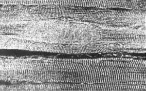
The working theory on how MTrPs form is called the Integrated Trigger Point Hypothesis when sarcomeres contract and increase the diameter of a muscle fiber forming a “contraction knot”. It is thought that this phenomenon occurs when muscle damage results in excessive release of acetylcholine at the motor endplate leading to sarcomere contracture. With this constant contracture, an energy crisis occurs due to muscle energy consumption exceeding the energy supply. This is thought to occur due to prolonged actin-myosin coupling, which leads to muscle fiber contraction and increased resistance to flow in the microvascular bed of the contracted muscle. This results in decreased oxygen and glucose levels, which in turn, reduces ATP production. Reduced ATP production may interfere with the reuptake of the Ca2+ into the sarcoplasmic reticulum which then prolongs the actin-myosin interaction and incites a vicious cycle. (See image 1)
How does this relate to veterinary patients with osteoarthritis?
It is common knowledge in human medicine that muscle pain, or myalgia, plays a big role in patients suffering from osteoarthritis. It is reported that MTrPs are more likely to be present when articular disease exists. We also understand that our patients suffering from osteoarthritis have common compensatory postural changes which lead to overuse of the affected muscles.
For example, patients suffering from osteoarthritis of the stifle will commonly be painful upon palpation of the cranial and caudal thigh (quadriceps and hamstrings). Patients with coxofemoral osteoarthritis often have increased pain in the gluteal and adductor muscles. Dogs that suffer from cranial cruciate injury and are not bearing weight on the injured limb typically have pain and muscle shortening of the iliopsoas, sartorius, and rectus femoris muscles of the ipsilateral limb. Any pelvic limb pain can cause the dog to shift excess weight to the thoracic limbs, increasing risk of overuse of the deltoids, triceps, infraspinatus and latissimus dorsi muscles. All of the muscles mentioned are common locations for MTrPs to be found.
How do you find MTrPs?
There are no validated laboratory tests or imaging techniques used to localize MTrPs. However, electromyography, ultrasonography, muscle biopsy, and thermography, have been studied. Thankfully, the best and least expensive way to find MTrPs is by palpation. However, careful muscle palpation is not something veterinarians are typically taught in school. It takes practice and understanding of proper palpation techniques.
First, to find the taut band, flat palpation with the pads of the finger perpendicular to muscle fiber alignment is needed. Going “against the grain” of the muscle allows for easier identification of the edge of the taut band. Once the taut band is located, follow parallel to the fibers until the contracted knot is found. MTrPs have been described as feeling like a grain of rice or marble in the muscle fibers. Additionally, a pincer technique may be used where an entire muscle group is grasped between the thumb and fingers and the muscle fibers are rolled between the tips of the digits to localize taut bands. This technique is useful to palpate the triceps or sartorius muscles.
What do you do after you locate an MTrP?
There are several reported methods of treatment in human medicine and many are being utilized in veterinary medicine as well.
The first treatment suggested by Travell and her colleague Simmons is termed ischemic compression. Ischemic compression is a manual therapy of MTrPs that consists of the application of sustained pressure for a long enough time (up to 90 seconds) to inactivate the point. Oftentimes, the therapist feels the MTrP give way under the pressure. The pressure on the point causes an increase in hypoxia, but as the pressure is released, blood rushes back into the area which helps to deactivate the MTrP. This method is non-invasive and well tolerated by our veterinary patients.
Dry needling is also a commonly utilized treatment method. A filiform acupuncture needle is placed into the point. Some practitioners will leave the needle in place for a period of time, while others will use a piston technique going in and out of the MTrP to help deactivate it. The application of the needle releases the sarcomere contracture and has been shown to produce a local twitch response (LTR) which provides immediate local pain relief to the area. Additionally, in humans, referred pain and widespread pain sensation are reduced with dry needling by reducing peripheral and central sensitization. Most veterinary patients require mild sedation for dry needling of the trigger points. Interestingly, the LTR will be present even in sedated patients.
Should you treat MTrPs?
A study performed on 48 chronically lame dogs with confirmed trigger points in some of the most common locations (triceps, infraspinatus, adductor, pectineus, peroneus longus, gluteus medius, iliocostalis lumborum, and quadriceps femoris) were treated weekly with either dry needling with a 28 gauge needle or injection of the trigger point with 1% lidocaine using a 23 gauge needle. Some patients were treated once, while others were treated up to 11 weeks. The dogs were evaluated for resolution of MTrPs as well as reduced lameness from 2 months to 6 years post-treatment. 60% of the patients demonstrated improvement. Some of these patients had received a variety of other treatments that failed to provide relief. Treating MTrPS should be an option in your chronic pain toolbox because this type of pain may not respond to traditional methods currently used by the majority of veterinarians.
Additionally, MTrPs have the ability to inhibit the muscle’s normal firing mechanism. When the muscle activation pattern is disrupted, muscle weakness is present. However, when MTrPs are deactivated by dry needling, in this author’s experience, an immediate return of strength is apparent in many patients. Muscles that are shortened due to constant contracture also reduce the range of motion of the joints they act upon. When the MTrPs are released, the joint range of motion improves as well.
Extracorporeal shockwave therapy (ESWT) is another treatment option for MTrPs. A 2019 study in humans revealed 3 treatments of radial ESWT provided as much pain relief as the dry needling technique. Photobiomodulation therapy (LASER therapy) has been evaluated with mixed results. Oral medications such as NSAIDs, tricyclic antidepressants, and muscle relaxants have also been evaluated with equivocal outcomes.
How can we prevent MTrPs?
Prevention of MTrPs is an interesting idea as well. If we can predict situations where MTrPs are likely to form, intervention ahead of time is plausible. In a randomized, double-blinded placebo-controlled study in humans looking at a single dry needle treatment of MTrPs immediately prior to total knee arthroplasty versus a sham group revealed the MTrPs treated group had significantly reduced pain based on visual analog scale (VAS) measurements 1 month after surgery.
The treatment group also required less pain medication intervention during the first month which is considered the most painful period during recovery. There was no lasting benefit at the 3 and 6-month rechecks. However, it is plausible that continued treatment of the MTrPs would provide a longer pain relief benefit during recovery periods.
This is an exciting concept to consider when looking at multimodal pain control options for our patients. Might pretreatment of MTrPs in our TPLO patients improve their pain during recovery? This is an area where further research is needed. Furthermore, mapping the referred pain pathways would be a great addition to our understanding of MTrPs in veterinary patients.
As our knowledge of myofascial pain grows, our treatment options for our patients suffering from chronic pain will expand as well. But before we can treat MTrPs, we must be able to recognize them and their clinical importance. Looking beyond the joints for pain and closely evaluating our patients’ muscles will provide us with clues to our patient’s condition and a path to more complete treatment of our canine companions.
- To learn more about Canine Myofascial Trigger Point Management, consider attending:
Myopain Seminars
July 10-11, 2020
The Woodlands, Texas.
Presented by Rick Wall, DVM, DACVSMR, CCRP, CMTPT
The course is open to veterinarians, physical therapists, and registered veterinary technicians.
For more information, click here.
References
Simons DG, Travell JG, Simons LS. Travell and Simons’ Myofascial Pain and Dysfunction: The Trigger Point Manual. Vol. 876. 1. 2nd ed. Baltimore, MD: Williams & Wilkins, 1999
Dommerholt J, Bron C, and Franssen J. Myofascial Trigger Points: An Evidence-Informed Review. The Journal of Manual & Manipulative Therapy. 2006 14:203-221
Wall R. Introduction to Myofascial Trigger Points in Dogs. Topics in Companion Animal Medicine, 29 (2014) 43-48
Janseens L. Trigger Points in 48 Dogs with Myofascial Pain Syndromes. Veterinary Surgery, 1991 20, 4: 274-278
Mayoral O, Salvat I, et al. Efficacy of Myofascial Trigger Point Dry Needling in the Prevention of Pain after Total Knee Arthroplasty: A Randomized, Double-Blinded, Placebo-Controlled Trial. Evid Based Complement Alternat Med 2013; 2013:694941 doi: 10.1155/2013/694941. Epub 2013 Mar 27
Yentür A, Okçu G. Yegül I. The role of trigger point therapy in knee osteoarthritis, The Pain Clinic, 2003 15:4, 385-390, DOI: 10.1163/156856903770196746
Luan S, Zhu Z, et al. Randomized Trial on Comparison of the Efficacy of Extracorporeal Shock Wave Therapy and Dry Needling in Myofascial Trigger Points. American Journal of Physical Medicine and Rehabilitation, 2019 98: 677-684 doi: 10.1097/PHM.0000000000001173
Kalichman L and Vulfsons S. Dry Needling in the Management of Musculoskeletal Pain. The Journal of the American Board of Family Medicine. 2010 23:640-646 doi.org/10.3122/jabfm.2010.05.090296
Alvarez D, Rockwell P. Trigger Points: Diagnosis and Management. Curr Pain Headache Rep. 2012 Oct; 16 (5):439-444 doi 10.1007/s11916-012-0289-4
Minerbi A, Vulfsons S. Challenging the Cinderella Hypothesis: A New Model for the Role of the Motor Unit Recruitment Pattern in the Pathogenesis of Myofascial Pain Syndrome in Postural Muscles. Rambam Maimonides Med J. 2018 Jul 9 (3): e0021