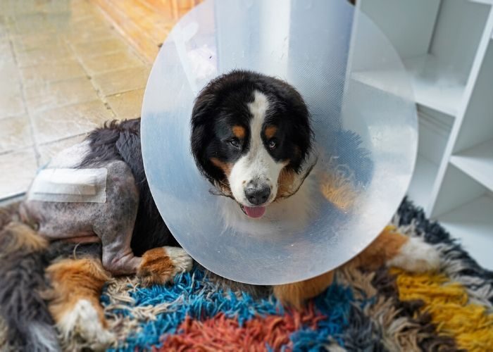
Hip Dysplasia/ Hip Arthritis
There are 4 types of surgical treatment for HD: two procedures are performed in juvenile dogs only with the goal of preventing the development of OA; two are typically performed in an adult dog that is symptomatic for OA.
- Juvenile Pubic Symphysiodesis (JPS): This is a relatively simple procedure that involves cauterizing the pubic symphysis in order to alter the growth of the acetabulum. When this is performed in puppies between 16-20 weeks of age, the development of the pelvis is improved so that the femoral head sits within the acetabulum and less arthritis can be expected long term. The JPS surgery is most effective in dogs with mild to moderate hip laxity and must be before 20 weeks of age; however, we do not recommend spaying or neutering client-owned dogs this young. Should a dog have this procedure performed, it is advised that the client sign a consent that they will not breed their dog as the surgery does not prevent passing the genetic trait of HD on to offspring.
- Triple or Double Pelvic Osteotomy (TPO/DPO): This surgery involves cutting the pelvis in 2 or 3 places and rotating the acetabulum to provide better coverage of the femoral head. This is a more invasive procedure than the JPS. It is performed in puppies typically between 6-8 months of age. It can be effective in reducing the progression of arthritis but only if it performed prior to the onset of visible arthritis (best assessed arthroscopically). Consultation with a surgeon is recommended to discuss this procedure further.
Treatment of arthritis secondary to HD is accomplished by either replacing the joint or removing the articulation and bone-on-bone contact. These surgeries are typically reserved for when non-surgical management efforts have been exhausted.
- Total Hip Replacement (THR): This is the gold standard surgery for HD, and results can be excellent with dogs returning to very active lives. However, there are potential risks associated with surgery. If non-surgical treatment of HD has been unsuccessful, consultation with a surgeon is recommended to discuss the pre-operative workup, post-operative care, and potential risks of THR.
- Femoral Head and Neck Ostectomy (FHO): This procedure removes the femoral head and neck, thus removing the bone-on-bone contact of the hip. Many dogs do well with this procedure, but post-operative rehabilitation soon after surgery is very important, especially for large dogs.
Legg-Calve Perthes (Avascular Necrosis of the Femoral Head)
The most common and successful treatment for LCP is femoral head and neck ostectomy (FHO). This surgery carries an an excellent prognosis, with reports of relieving pain and lameness in 80-100% of dogs. Rehabilitation is strongly recommended following FHO. Conservative/ non-surgical management is not recommended unless surgery is not feasible due to financial or medical limitations.
Elbow Dysplasia
Fragmented Medial Coronoid Process (FMCP)
FMCP is a type of elbow dysplasia that eventually leads to OA. There are several surgical options that may be considered for young dogs diagnosed with FMCP. These include:
- Arthroscopic fragment removal
- Subtotal coronoidectomy
- Variations of ulnar osteotomy or ostectomy
- Biceps ulnar release procedure (BURP).
While most surgeons report improvement in lameness following fragment removal and other procedures, surgery will not be curative and long term management of arthritis will be required.
Ununited Anconeal Process (UAP)
There are several surgical options that may be considered for young dogs diagnosed with UAP. These include:
- Arthroscopy to evaluate the entire joint
- Reattaching the anconeal process with a screw
- Ulnar osteotomy to relieve tension on the UAP so that it may fuse
- Excision of the anconeal process. Like other components of elbow dysplasia, UAP will likely result in some degree of long term OA.
Osteochondrosis/ Osteochondritis dissecans (OC/OCD)
Treatment for OCD consists of surgical (typically arthroscopic) debridement of the flap and abnormal cartilage. Then, the most common treatment is osteostixis, or the creation of small holes into the underlying subchondral bone to encourage in-growth of fibrocartilage (scar-tissue-type cartilage). Some surgeons will then inject PRP or other biologic therapies to encourage healing (though growth of normal hyaline cartilage has not been proven with these treatments).
Alternatively, a surgeon may elect to place an autologous, allogeneic or synthetic graft in the OCD defect. The autograph procedure is called Osteochondral Autograph Transfer System (OATS); the synthetic graft is called Synacart. Not all dogs with OC/OCD are candidates for these procedures, and long term results are mixed. Consultation with a local surgeon is advised to learn if this procedure is offered in your region.
Elbow Replacement
Unlike HD, joint replacement for elbow OA is not yet considered gold standard. There are surgeons working on refining the implants and surgical technique, but to date, total elbow replacement is not widely performed and carries a large number of risks.
Other surgical options for elbow arthritis in mature dogs include canine unicompartmental elbow replacement (CUE) and Sliding Humeral Osteotomy (SHO); these procedures are also not widely performed and have shown mixed results in clinical patients.
It is important to realize that while surgery often provides improvement in lameness in dogs with all forms of elbow dysplasia, surgery will not be a curative and long term management of arthritis will be required.
Cranial Cruciate Ligament Disease
While not traditionally considered to be a developmental orthopedic disease, cranial cruciate ligament injury is believed to be a degenerative condition with genetic and conformational influences.
Surgical stabilization of the stifle joint is the treatment of choice for CCL disease in dogs. The aim of surgery is to stabilize the joint and prevent the abnormal motion that occurs with cruciate insufficiency that leads to progressive arthritis and meniscal injury.
While surgery cannot prevent arthritis of the stifle (it is possible that underlying synovitis precedes CCL tearing), surgery can slow the progression of OA. Surgery is often recommended for partial tears of the CCL (Grade 1-2 sprain) as the ligament is not expected to heal and surgery can typically return a dog to an active lifestyle faster than non-surgical management.
Dogs are expected to return to their prior level of activity about 4 months after CCL surgery, whereas 12 months or longer is needed to achieve “good” results without surgery. If the meniscus is torn, arthritis will progress more rapidly and non-surgical management will be challenging.
There are several different surgical procedures that are broadly categorized into osteotomy (TPLO, TTA, CBLO) and extra-capsular procedures (Lateral suture, MRIT, Tight Rope). Studies so far support the TPLO as the procedure most likely to restore normal limb function. This is a great site for more information on TPLO: TPLOInfo.com
While surgery is typically the fastest way of returning dogs to an active lifestyle, it may not be the best option for every dog. Non-surgical management may be recommended in the following scenarios:
- Concurrent medical conditions that preclude anesthesia or surgery, examples include:
- Severe anemia
- Severe neurological conditions
- Kidney or liver failure
- Terminal cancer
There are no long-term studies showing that non-surgical management of CCL disease can guarantee that surgery is avoided, whereas there are many studies that document the progression of arthritis in a cruciate-deficient (unstable) stifle joint.
Patella Luxation
Surgical treatment is generally recommended for dogs with Grade III or IV MPL, frequent lameness associated with Grade II MPL, and animals with concurrent CCL rupture (MPL predisposes to CCL tearing).
Surgical procedures aim to realign the quadriceps-patellar mechanism. While surgery is tailored to the individual patient, it usually involves trochleaplasty (wedge or block), re-alignment of the tibial tuberosity (tibial tuberosity transposition, anti-rotational suture), and imbrication of the stretched tissues (ie, lateral imbrication for MPL). Distal femoral osteotomy/ostectomy may be recommended for dogs with significant distal femoral varus.
Patella luxation has been shown to lead to stifle OA, particularly if CCL rupture occurs. Therefore, long term management of OA is recommended.
References
Dueland RT, Adams WM, Patricelli AJ, et al. Canine hip dysplasia treated by juvenile pubic symphysiodesis. Part 1: Two year results of computed tomography and distraction index. VCOT 2010.
Dueland RT, Adams WM, Patricelli AJ, et al. Canine hip dysplasia treated by juvenile pubic symphysiodesis. Part 2: Two year clinical results. VCOT 2010.
Vezzoni A, Dravelli G, Vezzoni L, et al. Comparison of conservative management and juvenile pubic symphysiodesis in the early treatment of canine hip dysplasia. VCOT 2008.
Slocum B, Devine T. Pelvic osteotomy for axial rotation of the acetabular segment in dogs with hip dysplasia. Vet Clin North Am Small Anim Pract 1992.
Johnson AL, Smith CW, Pijanowski GJ, Hungerford LL. Triple pelvic osteotomy: effect on limb function and progression of degenerative joint disease. J Am Anim
Hosp Assoc. 1998 May-Jun;34(3):260-4.
Vezzoni A, Boiocchi S, Vezzoni L, et al. Double pelvic osteotomy for the treatment of hip dysplasia in young dogs. VCOT 2010.
Conzemius MG, Vandervoort J. Total joint replacement in the dog. Vet Clin North Am Small Anim 2005
Piek CJ, Hazewinkel HA, Wolvekamp WT, et al. Long-term follow-up of avascular necrosis of the femoral head in the dog. J Small Anim Pract 1996 37(1):12-18.
Harper TAM. Femoral head and neck excision. Vet Clin North Am Small Anim Pract 2017.
Krotscheck U, Hulse DA, Bahr A, Jerram RM. Ununited anconeal process: lag-screw fixation with proximal ulnar osteotomy. V Comp Ortho Trauma 13:212-216.
Pettitt RA, Tattersall J, Gemmill T, et al. Effect of surgical technique on radiographic fusion of the anconeus in the treatment of ununited anconeal process. J Small Anim Prac 2009 50:545-548.
Evans RB, Gordon-Evans WJ, Conzemius MG. Comparison of three methods for the management of fragmented medial coronoid process in the dog: A systematic review and meta-analysis. Vet Comp Orthop Traumatol 2008 21:106-109.
Burton NJ, Owen MR, Kirk LS, et al. Conservative versus arthroscopic management for medial coronoid process disease in dogs: A prospective gait evaluation. Vet Surg 2011 40:972-980.
Fitzpatrick N, Yeadon R, Smith T, Schulz K. Techniques of application and initial clinical experience with sliding humeral osteotomy for treatment of medial compartment disease of the canine elbow. Vet Surg 2009 38:261-278.
Conzemius M. Nonconstrained elbow replacement in dogs. Vet Surg 2009 38:279-284.
Fitzpatrick N, Yeadon R, Smith TJ. Early clinical experience with osteochondral autograft transfer for treatment of osteochondritis dissecans of the medial humeral condyle in dogs. Vet Surg 2009 38:246-260.
Slocum B, Slocum T. Tibial plateau leveling osteotomy for repair of cranial cruciate ligament rupture in the canine. Vet Clin N Am-Small 23:777–95, 1993
Hoffmann DE, Miller JM, Ober CP, et al: Tibial tuberosity advancement in 65 canine stifles. Vet Comp Orthop Traumatol 19:219–227, 2006
Kim SE, Pozzi A, Kowaleski MP, et al: Tibial osteotomies for cranial cruciate ligament insufficiency in dogs. Vet Surg 37:111–125, 2008
Raske M, Hulse D, Beale B, et al: Stabilization of the CORA based leveling osteotomy for treatment of cranial cruciate ligament injury using a bone plate augmented with a headless compression screw. Vet Surg 42: 759-764, 2013.
Gordon-Evans WJ, Griffon DJ, Bubb C, et al: Comparison of lateral fabellar suture and tibial plateau leveling osteotomy techniques for treatment of dogs with cranial cruciate ligament disease. J Am Vet Med Assoc 243:675-680, 2013
Wucherer KL, Conzemius MG, Evans R et al: Short-term and long-term outcomes for overweight dogs with cranial cruciate ligament rupture treated surgically or nonsurgically. J Am Vet Med Assoc 2013;242:1364-1372
L’Eplattenier H, Montavon P. Patellar luxation in dogs and cats: Management and prevention. Compend 2002 24(4)292-299.
Johnson AL, Probst CW, Decamp CE, et al. Comparison of trochlear block recession and trochlear wedge recession for canine patellar luxation using a cadaver model. Vet Surg 2001 30:140-150.
Langenbach A, Marcellin-Little DJ. Management of concurrent patellar luxation and cranial cruciate ligament rupture using modified tibial plateau leveling. J Small Anim Prac 2010 51:97-103.
Yeadon R, Fitzpatrick N, Kowaleski MP. Tibial tuberosity transposition-advancement for treatment of medial patellar luxation and concomitant cranial cruciate ligament disease in the dog. V Comp Orthop Traumatol 2011 24:18-26.